See a GP if you're concerned about your child's weight. Measuring waist size is not routinely recommended for children because it does not take their height into account.

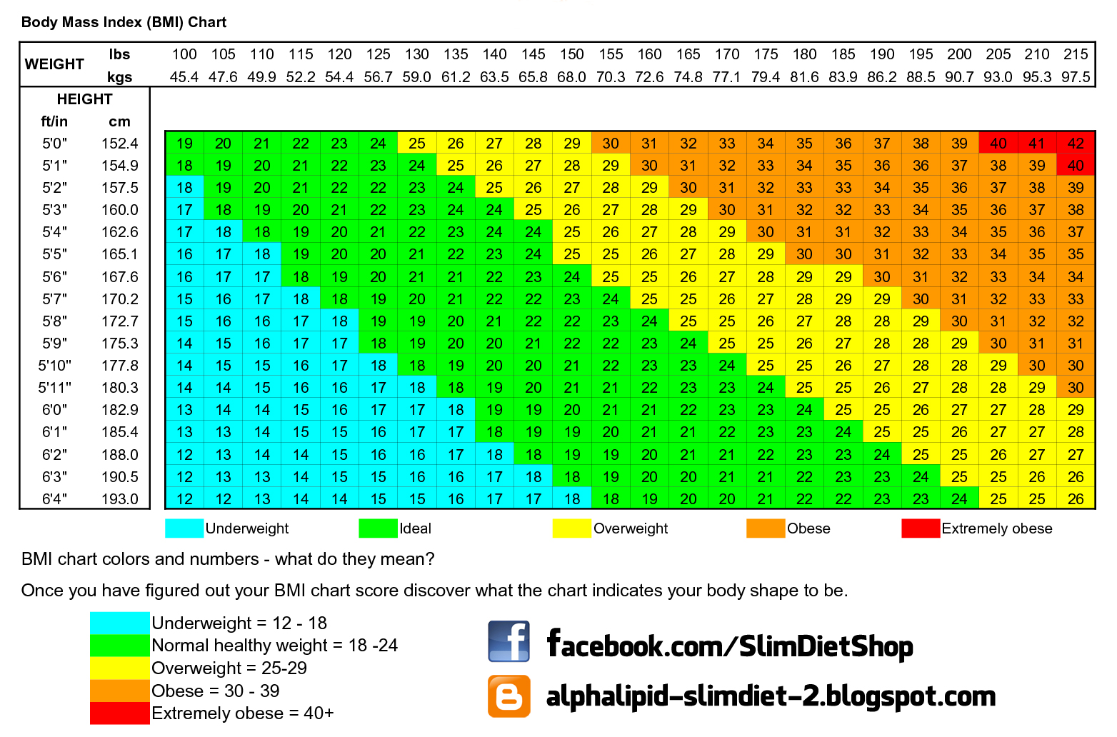
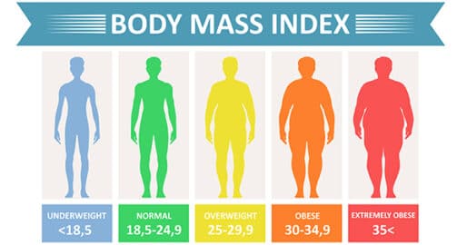
Breathe out naturally before taking the measurement.Wrap a tape measure around your waist midway between these points.Find the bottom of your ribs and the top of your hips.You can have a healthy BMI and still have excess tummy fat, meaning you're still at risk of developing these conditions. Measuring your waist is a good way to check you're not carrying too much fat around your stomach, which can raise your risk of heart disease, type 2 diabetes and stroke. Black, Asian and other minority ethnic groupsīlack, Asian and other minority ethnic groups have a higher risk of developing some long-term (chronic) conditions, such as type 2 diabetes. The best way to lose weight if you're obese is through a combination of diet and exercise, and, in some cases, medicines. The BMI calculator will give you a personal calorie allowance to help you achieve a healthy weight safely. The best way to lose weight if you're overweight is through a combination of diet and exercise. Keep up the good work! For tips on maintaining a healthy weight, check out the food and diet and fitness sections. If you're underweight, a GP can help.įind out more in underweight adults Healthy weight 2003 17(1):31-39.Understanding your BMI result Underweightīeing underweight could be a sign you're not eating enough or you may be ill. Left ventricular mass index measured by quantitative gated myocardial SPECT with 99mTc-tetrofosmin: a comparison with echocardiography. Maruyama K, Hasegawa S, Nakatani D, et al. Left ventricular relative wall thickness versus left ventricular mass index in non-cardioembolic stroke patients. Hashem MS, Kalashyan H, Choy J, Chiew SK, Shawki AH, Dawood AH, Becher H. Normal Values of Left Ventricular Mass Index Assessed by Transthoracic Three-Dimensional Echocardiography. Mizukoshi K, Takeuchi M, Nagata Y, et al. Standardization of adult transthoracic echocardiography reporting in agreement with recent chamber quantification, diastolic function, and heart valve disease recommendations: an expert consensus document of the European Association of Cardiovascular Imaging. Recommendations for cardiac chamber quantification by echocardiography in adults: an update from the American Society of Echocardiography and the European Association of Cardiovascular Imaging. Relative wall thickness (RWT) allows classification of LV mass increase as either:

Individuals with LV hypertrophy were found to be at 2.7 times greater risk for cardiac events, such as myocardial infarction or coronary heart disease death, (over 15 years) than individuals without LV hypertrophy. Left ventricular mass (LVM) can be directly determined using cardiac magnetic resonance (CMR) and is known to increase in proportion to overall body size and differs by gender: Female

LVEDD: Left ventricular end-diastolic dimension.The parameters involved are summarised below:

Left ventricular mass is an important determinant of diagnosis and prognosis in patients with heart disease (cardiovascular morbidity and mortality) in specific for determination of severity and type of cardiac hypertrophy.


 0 kommentar(er)
